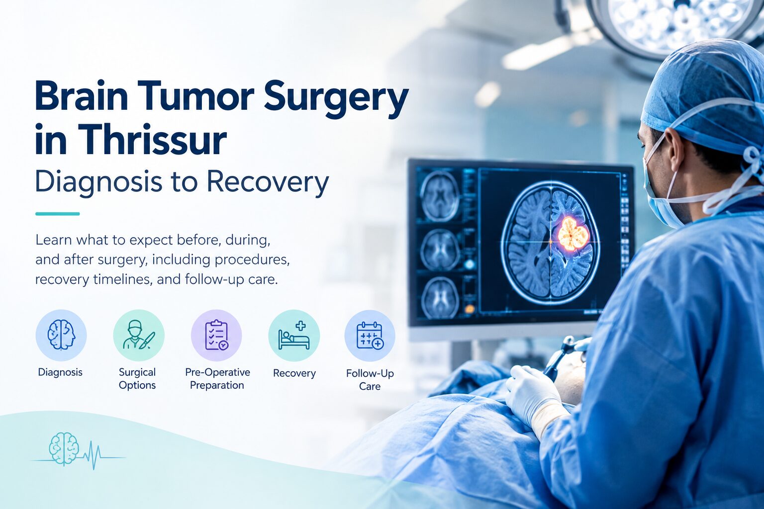
A brain tumor diagnosis changes the tempo of everything. Between the first suspicious scan and the first day back at work, most families in Thrissur move through a period of intense decision-making with very little clarity about what happens at each step. This guide walks through the full arc of brain tumor surgery, from the imaging that confirms the diagnosis to the weeks and months of recovery afterward. It reflects how the neurosurgery team at Atreya Hospital, Thrissur, approaches each stage.
How Brain Tumors Are Diagnosed
A brain tumor is rarely the first thing a doctor suspects. Most patients arrive at a neurology consultation after weeks or months of symptoms that gradually became harder to ignore. The diagnostic process combines a careful clinical assessment with high-resolution imaging.
Symptoms That Prompt a Neurology Consultation
The most common presenting complaints include:
- Persistent headaches, often worse in the morning or that wake the patient from sleep
- New-onset seizures in an adult who never had them before
- Progressive vision changes, double vision, or loss of peripheral vision
- Unexplained memory lapses, personality changes, or confusion
- Difficulty with speech, word-finding, or comprehension
- Weakness, numbness, or loss of coordination on one side of the body
- Persistent nausea or vomiting without a clear cause
A single symptom in isolation is rarely a tumor. A combination that develops over weeks or worsens progressively warrants specialist evaluation.
The Neurological Examination
A neurologist assesses cranial nerve function, muscle strength, reflexes, coordination, gait, and cognitive performance. This examination often identifies which part of the brain is affected before any imaging is done. It also establishes a baseline that later scans can be compared against.
MRI and CT Imaging
MRI with contrast is the primary diagnostic tool. It provides high-resolution images of soft tissue and shows tumor size, location, borders, and its relationship to surrounding structures. Most tumors are first identified or ruled out on MRI.
CT scans are often used first in emergency settings because they are faster and more widely available. CT is good at detecting bleeding, calcification, and bone involvement, but MRI is required for surgical planning in nearly all cases.
Advanced Imaging When Needed
For tumors near critical brain areas or major blood vessels, additional studies may be ordered. MR spectroscopy analyzes the tumor’s chemical composition and helps distinguish tumor from other conditions such as infection or stroke. Functional MRI maps speech and motor areas so the surgeon can plan around them. A cerebral angiogram is used when a tumor is close to major blood vessels or when a vascular malformation is suspected.
Confirming the Tumor Type
Not every brain lesion is a tumor, and not every tumor is cancerous. Roughly two-thirds of brain tumors diagnosed in adults are benign, meaning they do not spread. But benign does not mean harmless. A benign tumor pressing on critical brain tissue can still cause serious neurological symptoms and often needs surgical removal.
Once imaging suggests a tumor, a biopsy or the surgery itself provides the tissue sample that confirms the exact type. Meningiomas, gliomas, pituitary adenomas, schwannomas, and metastatic tumors from cancers elsewhere in the body all require different surgical strategies and post-operative care plans. Patients in Thrissur can access the full diagnostic workup, including advanced MRI and neurological assessment, at Atreya Hospital’s neurology department, which coordinates with the neurosurgery team to arrive at an accurate diagnosis before any surgical decision is made.
What Are the Types of Brain Tumor Surgery
Brain tumor surgery is not one procedure. The right surgical approach depends on the tumor’s type, location, size, and proximity to functional brain areas. The neurosurgery team selects the approach after multidisciplinary review.
1. Craniotomy
Craniotomy is the standard approach for most brain tumors. The surgeon temporarily removes a section of the skull to access the tumor, resects as much of it as safely possible, and then replaces the bone. Modern craniotomies use image-guided navigation to plan the smallest possible opening, which reduces trauma to surrounding tissue and shortens recovery.
2. Awake Craniotomy
Awake craniotomy is used when a tumor sits near areas that control speech, language, or movement. The patient is sedated for the opening and closing of the skull but kept awake during tumor removal itself. This allows the surgical team to test speech and motor function in real time, so the surgeon can maximize tumor removal without damaging critical function. The procedure sounds daunting, and it is a serious operation, but the awake portion is designed to be pain-free and closely supervised.
3. Endoscopic Surgery
Endoscopic surgery is often used for tumors of the pituitary gland or those inside the fluid-filled ventricles of the brain. The surgeon uses a thin endoscope, sometimes passed through the nose for pituitary tumors, avoiding a traditional skull opening altogether. Recovery times are typically shorter, and there are no visible scars.
4. Minimally Invasive Tumor Surgery
Minimally invasive tumor surgery uses smaller incisions and specialized retractors to reach deep-seated tumors through narrow corridors. When combined with intraoperative navigation and microscopic visualization, this approach can access lesions that were previously considered inoperable. It is not always appropriate, but where it is, patients typically experience less pain and faster recovery.
5. Stereotactic Biopsy
Stereotactic biopsy is a diagnostic rather than curative procedure. A thin needle is guided to the tumor using imaging coordinates, and a small tissue sample is taken for pathological analysis. This is used when a tumor is in a location too risky to remove completely, or when the tumor type needs to be confirmed before deciding on further treatment such as radiation or chemotherapy.
The Atreya neurosurgery team performs the full range of these procedures, including awake craniotomies, endoscopic surgery for pituitary and intraventricular tumors, and pediatric brain tumor surgery. Complex cases are reviewed through the Brain Tumor Clinic, which meets weekly for consultations and holds a monthly tumor board for multidisciplinary planning.
What Happens Before Brain Tumor Surgery
The period between diagnosis and surgery usually spans one to three weeks, longer if the tumor is slow-growing and there is time for careful planning. This window is used to complete pre-operative assessments and to prepare the patient physically and mentally.
1. Pre-Operative Workup
Standard pre-operative assessments include:
- An updated MRI closer to the surgery date, sometimes with functional or spectroscopy sequences
- Blood tests covering complete blood count, coagulation profile, kidney and liver function, and blood group
- A chest X-ray and an ECG
- An anesthesia consultation to review medical history and plan the anesthetic
- Cardiac or endocrine review for patients with diabetes, hypertension, or heart disease
- A neuropsychological evaluation in selected cases, to establish a cognitive baseline
Most of this workup can be completed on an outpatient basis over three to five days before admission.
2. Second Opinions and the Tumor Board
A second opinion is reasonable and often welcomed by the surgical team. Brain tumor surgery is one of the most consequential decisions a patient will make, and reviewing the plan with a second neurosurgeon can bring clarity and confidence.
At Atreya Hospital, complex cases are routinely discussed in a tumor board, where neurosurgeons, neurologists, radiologists, and oncologists review imaging together and agree on the best approach. This is standard practice at accredited neuro centres and should be expected, not requested.
3. Medication Adjustments
Medication changes are common in the days before surgery. Blood-thinning medications such as aspirin or clopidogrel usually need to be stopped several days in advance. Anti-seizure medication may be started prophylactically. Corticosteroids are often prescribed to reduce swelling around the tumor. The surgical team provides a specific list of what to stop, what to continue, and when.
4. The Night Before Surgery
The night before surgery, patients fast for six to eight hours before anesthesia. A section of the scalp is shaved at the surgical site, though modern practice preserves as much hair as possible. Family members are counselled about hospital stay expectations, ICU protocols, and what to bring.
5. Emotional Preparation
Emotional preparation matters as much as physical preparation. Patients who understand the procedure, the risks, and the expected recovery generally cope better afterward. The pre-op consultation is the time to ask about surgery duration, tumor-specific risks, and what recovery looks like at one week, one month, and six months. A good surgical team welcomes these questions and answers them without hurry.
What Happens During Brain Tumor Surgery
The operating room for a brain tumor procedure is a highly technical environment. Understanding what happens inside it can reduce a lot of anxiety for families waiting outside.
1. Anesthesia and Positioning
After the patient is brought into the operating theatre, general anesthesia is administered by a neuroanesthesia team. The head is then positioned and secured in a rigid frame that prevents any movement during surgery. The angle of approach affects how safely the tumor can be reached, so positioning is done with care.
2. Neuronavigation Setup
The surgical site is cleaned, and the incision is planned using pre-operative MRI images that are registered to the patient’s anatomy through a neuronavigation system. Neuronavigation works like a GPS for the brain, giving the surgeon real-time three-dimensional guidance on where the tumor sits relative to critical structures such as the motor cortex, speech areas, and major blood vessels.
3. Intraoperative Microscopy and Fluorescence
Once the skull is opened and the dura (the tough membrane covering the brain) is carefully incised, the tumor is approached through the safest available corridor. At Atreya Hospital, the neurosurgery operation theatre is equipped with the Carl Zeiss Kinevo 900 microscope, which provides high-magnification, high-definition visualization of tumor tissue and surrounding structures. The microscope includes a yellow filter that visualizes fluorescent-marked tumor tissue during surgery, helping the surgeon distinguish tumor from healthy brain more precisely. For tumors near major blood vessels, intraoperative angiography using ICG dye allows the surgeon to see blood flow in real time and avoid vascular injury.
4. Intraoperative Neuromonitoring
Intraoperative neuromonitoring (IONM) continuously tracks motor and sensory pathways throughout the procedure. Small electrodes placed on the scalp and body monitor the brain’s electrical responses. If any surgical action risks affecting a critical function, the monitoring team alerts the surgeon immediately. IONM has significantly reduced the risk of permanent neurological deficits in modern brain tumor surgery.
5. Tumor Removal and Closure
Tumor removal is done in stages. The surgeon carefully dissects tumor tissue away from healthy brain, working under microscopic guidance and often using ultrasonic aspiration to break up and remove the tumor without pulling on surrounding structures. The goal is maximum safe resection: removing as much tumor as possible without compromising neurological function. In some cases, complete removal is not possible because of the tumor’s location, and residual tumor is treated with radiation or chemotherapy afterward.
Once tumor removal is complete, the dura is repaired, the bone flap is replaced and secured with titanium plates or sutures, and the scalp is closed in layers. Total surgery time typically ranges from three to six hours, though complex tumors can take longer. The patient is then transferred to the neuro ICU for close monitoring.
Recovery After Brain Tumor Surgery: Week by Week
Recovery from brain tumor surgery is a gradual process, not a single event. Understanding the typical timeline helps families plan for the weeks and months ahead.
First 24 to 48 Hours: Neuro ICU
The patient is monitored continuously in a neurosurgical intensive care unit. Vital signs, neurological status, and intracranial pressure are checked frequently, often every hour in the first day. Pain is managed with medication, and swelling is controlled with corticosteroids. Most patients are alert enough to speak and follow commands within a few hours of surgery, though drowsiness and disorientation are normal.
Days 3 to 7: Hospital Ward
Most patients transfer from ICU to a regular ward within one to three days if there are no complications. Early mobilization begins, often within 24 to 48 hours of surgery, because getting up and walking reduces the risk of blood clots and pneumonia. Diet is advanced gradually, starting with clear fluids and moving to a normal diet as tolerated. Stitches or staples are usually removed around day seven to ten. A post-operative MRI is done within 48 to 72 hours to assess the extent of tumor removal.
Weeks 1 to 4 at Home
Fatigue is the dominant symptom in the first month. This is normal and expected. The brain uses significant energy to heal, and most patients need frequent rest throughout the day. Headaches are common in the first two weeks and usually respond to prescribed medication. Physical activity should be limited to short walks. Driving, heavy lifting, and returning to work are not permitted during this period. Wound care instructions are provided at discharge.
Weeks 4 to 12: Gradual Return to Activity
Energy gradually returns through the second and third months. Patients typically resume desk-based work between four and six weeks after surgery, though full stamina takes longer. Driving may be permitted around six to eight weeks, depending on whether the patient had seizures and how the follow-up MRI looks. Physically demanding work often requires eight to twelve weeks. Physiotherapy, occupational therapy, or speech therapy is prescribed if the surgery affected specific brain regions.
Beyond Three Months: Long-Term Follow-Up
For benign tumors that were completely removed, follow-up imaging every six to twelve months monitors for recurrence. For malignant tumors, adjuvant treatment such as radiation or chemotherapy typically begins four to six weeks after surgery and continues over subsequent months. Rehabilitation for any residual neurological deficits continues as needed. Most patients return to their normal life activities by six months, though the exact trajectory varies by age, tumor type, and extent of surgery.
Patients in Thrissur benefit from having follow-up imaging, physiotherapy, and specialist review available locally, without repeated travel to Kochi or Bangalore.
Conclusion
A brain tumour diagnosis is not the end of the story. It is the beginning of a treatment path that, more often than not, leads to meaningful recovery and a return to the life you love. What matters most in these early days is finding a team that listens, explains, and treats you as a whole person.
Still Unsure About The Diagnosis?
If you or someone in your family is navigating the possibility of brain tumour surgery, the specialists at Atreya Hospital’s Brain Tumour Clinic in Thrissur are here to talk it through with you. You can book a consultation on Mondays between 10 AM and 1 PM, or reach out for appointments any day of the week.
To book an appointment, call +91 9387668030 or use the link below. Patients based outside Kerala, including in the GCC, can begin with a teleconsultation before travelling for care.