You pick up your spine MRI report, scan the findings section, and immediately run into words like “disc desiccation,” “broad-based disc bulge,” “annular fissure,” and “foraminal narrowing.” None of it sounds good. Most of it sounds alarming. And your follow-up appointment with the doctor is still a few days away.
This is one of the most common experiences patients describe after a spine MRI. The terminology in a radiology report is written for the referring physician, not for the person whose spine was scanned. That gap between what the report says and what it actually means for your health creates unnecessary anxiety.
This guide explains the most frequently used terms in a spine MRI report in plain language. It covers what each finding means, when it matters clinically, and what to expect when you bring the report to a spine specialist.
How a Spine MRI Works
MRI (magnetic resonance imaging) uses strong magnetic fields and radio waves to produce detailed images of the soft tissues in and around your spine. Unlike an X-ray, which shows bones well but soft tissues poorly, an MRI reveals the discs between your vertebrae, the spinal cord, nerve roots, ligaments, and surrounding muscles.
A spine MRI is typically ordered when a patient has persistent back or neck pain, pain radiating into the arms or legs (radiculopathy), numbness, tingling, weakness, or difficulty with balance or coordination. The scan helps the doctor see whether a structural problem in the spine could explain these symptoms.
Your MRI report covers one of three regions, sometimes more than one: the cervical spine (neck), the thoracic spine (mid-back), or the lumbar spine (lower back). The lumbar region is the most commonly scanned, since lower back pain is the most frequent reason for referral.
Understanding the Structure of Your Spine MRI Report
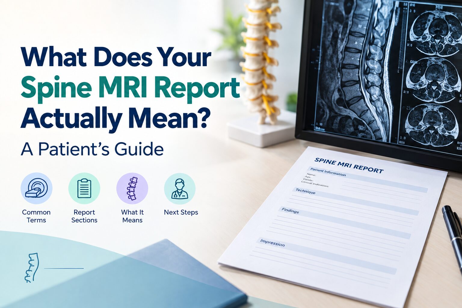
Every spine MRI report follows a standard format. Knowing the sections helps you locate the information that matters most.
- Patient information and clinical indication
The top of the report lists your name, age, and the reason your doctor ordered the scan (for example, “lower back pain with left leg radiculopathy”). This matters because the radiologist interprets the images with this clinical context in mind. - Technique
This section describes how the scan was performed: the type of MRI sequences used (T1-weighted, T2-weighted, STIR), whether contrast dye was administered, and which planes (sagittal, axial, coronal) were imaged. You generally do not need to interpret this section yourself. - Findings
This is the most detailed section. The radiologist describes what each vertebral level looks like, working through the discs, bones, spinal canal, nerve roots, and surrounding soft tissues. Most of the confusing terminology sits here. - Impression
The final section is a summary. It highlights the most significant findings and may suggest a possible diagnosis or recommend additional imaging. If you are short on time before your appointment, read the impression section first.
Common Terms in Your Spine MRI Report Explained
The terms below appear in the vast majority of spine MRI reports. Many of them describe age-related changes that are common in healthy adults over 30 to 35, and most do not automatically mean you need surgery or even treatment. Understanding each term puts you in a better position to have a meaningful conversation with your doctor.
1. Disc Desiccation
Disc desiccation means the disc between two vertebrae has lost some of its water content. On a T2-weighted MRI image, a healthy disc appears bright white because it is well-hydrated. A desiccated disc looks darker. This is one of the earliest visible signs of normal disc aging and is extremely common after the age of 30. Disc desiccation alone is usually not the cause of back pain. It becomes relevant when it is accompanied by other changes, such as a loss of disc height, a bulge, or a herniation.
2. Disc Bulge
A disc bulge means the disc extends outward beyond the edge of the vertebral body in a broad, generalized way, typically involving more than 25% of the disc’s circumference. The outer wall of the disc (the annulus fibrosus) remains intact. Think of it as a tire that has lost some air pressure and flattened slightly, causing the rubber to spread outward evenly.
Disc bulges are among the most commonly reported MRI findings. Research published in the American Journal of Neuroradiology has shown that disc bulges are found in a significant percentage of people over 40 who have no back pain at all. A disc bulge on your report does not, by itself, mean that the disc is causing your symptoms. It becomes clinically significant when it is large enough to narrow the spinal canal or compress a nerve root, and when that compression matches the pattern of your symptoms.
3. Disc Herniation (Protrusion and Extrusion)
A disc herniation is a more focal and more severe form of disc displacement than a bulge. In a herniation, the disc material pushes outward through a tear or weakness in the outer annulus, rather than spreading broadly.
Radiologists classify herniations into subtypes:
- Protrusion: The base of the herniation is wider than the part that extends into the spinal canal. The disc material has pushed out but remains connected to the main disc body.
- Extrusion: The herniated material has a narrower base than its displaced portion, meaning a piece of disc has pushed further out and may be separating.
- Sequestration: A fragment of disc material has broken off entirely and is sitting free in the spinal canal. This is the least common type but often the most symptomatic.
The location of the herniation also matters. Reports describe it as central (midline), paracentral (slightly off-center), foraminal (in the nerve exit channel), or far lateral (outside the foramen). A paracentral or foraminal herniation is more likely to compress a specific nerve root and cause pain radiating down a leg or arm.
4. Annular Tear (Annular Fissure)
An annular tear is a small tear or crack in the outer ring (annulus fibrosus) of the disc. On an MRI, it may appear as a bright spot on T2-weighted images, sometimes called a “high-intensity zone” (HIZ). Annular tears are very common and frequently asymptomatic. They can cause localized back pain in some cases, and they may progress over time into a disc herniation if the inner disc material pushes through the tear. Most annular tears heal on their own and do not require surgical intervention.
5. Spinal Stenosis
Spinal stenosis means the spinal canal (the channel through which the spinal cord and nerve roots pass) has narrowed. The narrowing can result from a combination of disc bulges, bone spurs (osteophytes), thickened ligaments, and enlarged facet joints. Radiologists grade stenosis as mild, moderate, or severe based on how much of the canal space is reduced.
Mild stenosis is common in adults over 50 and is often asymptomatic. Moderate to severe stenosis, particularly in the lumbar region, can cause symptoms like leg pain, heaviness, or cramping when walking (a pattern called neurogenic claudication), and may require treatment.
6. Neural Foraminal Narrowing
The neural foramina are small channels on either side of the spine where the nerve roots exit the spinal canal. When these channels narrow (due to disc bulges, bone spurs, or facet joint enlargement), the nerve passing through can become compressed. This is called foraminal stenosis or neural foraminal narrowing.
If the narrowing matches the nerve distribution that corresponds to your symptoms (for instance, foraminal narrowing at L5-S1 on the left side with pain radiating down the left leg), it is likely to be clinically significant. If it does not match, it may be an incidental finding.
7. Spondylosis and Degenerative Changes
Spondylosis is a general term for age-related wear and tear of the spine, including disc degeneration, bone spur formation, and facet joint arthritis. You may also see phrases like “degenerative disc disease” or “multilevel degenerative changes.” These terms describe the natural aging process of the spine and are found on imaging in most adults over the age of 40, regardless of symptoms.
Spondylosis is not a disease in the way a fracture or tumor is a disease. It is a description of structural aging. Treatment depends entirely on whether the degenerative changes are producing symptoms such as pain, nerve compression, or instability.
8. Modic Changes
Modic changes refer to signal changes in the bone marrow of the vertebral bodies adjacent to a degenerating disc. They are classified into three types:
| Type | What It Shows | What It Suggests |
| Modic Type 1 | Bone marrow edema (bright on T2, dark on T1) | Active inflammation near a degenerating disc; may be associated with back pain |
| Modic Type 2 | Fatty replacement of bone marrow (bright on both T1 and T2) | Chronic, stable degenerative change; less likely to cause active pain |
| Modic Type 3 | Sclerotic bone (dark on both T1 and T2) | End-stage degeneration; relatively uncommon |
Modic Type 1 changes are the most clinically relevant because they indicate active inflammation and are more commonly associated with pain at that spinal level.
9. Ligamentum Flavum Thickening
The ligamentum flavum is a ligament that runs along the back of the spinal canal. With age or repeated mechanical stress, it can thicken. A normal ligamentum flavum measures less than 4 mm. When it thickens beyond this, it can contribute to spinal stenosis by taking up space inside the canal. This is one of the most common contributors to lumbar spinal stenosis in older adults.
These two terms appear on nearly every spine MRI report, and they are the most frequently confused. The difference is structural.
Disc Bulge vs. Disc Herniation: Understanding the Difference
| Feature | Disc Bulge | Disc Herniation |
| Shape of displacement | Broad and generalized (more than 25% of circumference) | Focal and localized (less than 25% of circumference) |
| Outer disc wall (annulus) | Intact, stretched but not torn | Torn or ruptured |
| Severity | Usually mild; often age-related | Ranges from mild (protrusion) to severe (extrusion, sequestration) |
| Nerve compression | Uncommon unless the bulge is large | More likely, especially with paracentral or foraminal herniations |
| Symptoms | Often none | More often associated with radiating arm or leg pain |
The key takeaway: a bulge is almost always a milder finding than a herniation. Many disc bulges are incidental and require no treatment. A herniation, particularly one that compresses a nerve root and matches your symptoms, is more likely to need clinical attention.
When Spine MRI Findings Need Medical Attention
Most findings on a spine MRI report are incidental or age-related and do not require surgery. However, certain combinations of findings and symptoms should prompt timely specialist evaluation.
Symptoms that warrant prompt attention:
- Severe or worsening pain that radiates into a specific leg or arm and does not respond to medication
- Numbness or tingling that follows a specific nerve pattern (for instance, the outer calf and foot)
- Muscle weakness in a leg or arm, such as difficulty lifting the foot while walking (foot drop)
- Loss of bladder or bowel control, or a sudden inability to urinate. This combination with saddle-area numbness (inner thighs, buttocks) may indicate cauda equina syndrome, a surgical emergency
- Balance difficulties, clumsiness with fine motor tasks (buttoning a shirt, holding objects), or an unsteady gait, which may indicate spinal cord compression in the cervical region
MRI findings that are more likely to be clinically significant:
- Disc extrusion or sequestration compressing a nerve root
- Moderate to severe spinal stenosis, particularly with cord compression or myelopathy signals
- Modic Type 1 changes at the level matching your pain
- High-grade foraminal narrowing that correlates with radiculopathy symptoms
A spine surgeon or neurologist evaluates the MRI alongside your symptoms, physical examination, and neurological testing. An MRI finding alone, no matter how dramatic it reads on paper, does not determine treatment. The clinical picture as a whole does.
What to Do After You Receive Your Spine MRI Report
Reading the report before your follow-up visit is natural, and having a basic understanding of the terms helps you ask better questions. Here are practical steps to take between receiving your report and seeing your doctor.
- Bring the full imaging, not just the printed report
The written report is the radiologist’s interpretation, but your spine specialist needs to see the actual MRI images (typically provided on a CD or accessible through a digital portal). Many clinical decisions depend on details visible in the images that are not fully captured in the written report. - Write down your questions
Note the terms or findings that concern you most. Ask your specialist to explain which findings correlate with your symptoms and which are incidental. Not every finding on the report requires treatment. - Do not self-diagnose from the report alone
An MRI is an anatomical snapshot of your spine at one moment in time. It does not measure pain, inflammation levels, or your functional capacity. Two patients with identical MRI findings can have completely different symptom profiles. The report is one piece of the diagnostic puzzle, not the full picture. - Ask about next steps
Depending on the findings and your symptoms, your specialist may recommend conservative management (physical therapy, medications, activity modification), further diagnostic testing (nerve conduction studies, CT myelogram), or, in select cases, a surgical opinion.
Conclusion
Receiving a spine MRI report full of unfamiliar medical terms can feel overwhelming. But understanding what those terms actually mean often reveals something reassuring: many findings are common, age-related changes that do not automatically signal something serious, and the vast majority of patients with spine conditions do not require surgery.
What matters most is how your findings relate to your specific symptoms, your lifestyle, and your goals. That interpretation requires a skilled spine specialist.
Don’t Wait For The Emergency. Book A Visit Today
At Atreya Hospital’s Spine Clinic in Thrissur, our multidisciplinary team of spine surgeons, physiotherapists, and rehabilitation specialists takes exactly this approach. We combine advanced diagnostic imaging, experienced clinical assessment, and evidence-based treatment to give you a clear, personalised answer to one question: what does this mean for you?
Whether you need a second opinion on a confusing report, expert management for a disc problem, or advanced spine surgery, our NABH-accredited team is here to guide you at every step.
To book an appointment, call +91 9387668030 or use the link below. Patients based outside Kerala, including in the GCC, can begin with a teleconsultation before travelling for care.
Medical Disclaimer: The information provided in this article is intended for general informational purposes only and should not be considered a substitute for professional medical advice, diagnosis, or treatment. Always consult a qualified healthcare provider regarding any medical condition or treatment. If you have specific questions about your health, please consult your doctor or visit Atreya Hospital.